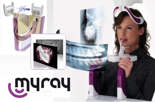
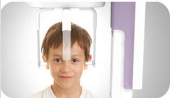
Hyperion features
Morphology Recognition Technology (MRT) which automatically
identifies patient size and all parameters required to
ensure correct X-ray exposure.
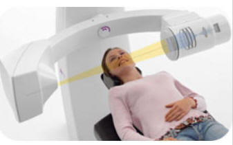
Allow your patient to
stand still, while the laser-guided multi-motor
kinematics positions itself in a matter of
seconds.
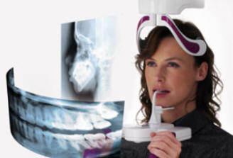
The face-to-face approach makes it
comfortable for both dentist and patient.
Reassuring eye contact with your patient is
possible at all times.
A fast scan reduces
possibility of patient movement. 7.9 seconds for
the child panoramic and just 9.2 seconds the
adult panoramic.
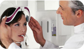
Failsafe procedures
Via laser-guided
multi-motor kinematics, Hyperion positions
itself around your patient, reads the subject
and scans accordingly.
No standardized single
rotation axis. Each exam is personalized to give
you utmost diagnostic precision.
With MRT
technology there is no need to program exposure
times or technical factors such as kV or mA
levels, nor is it necessary to select patient
build. Hyperion predicts your needs exactly.
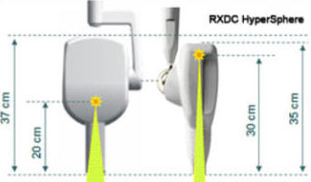
X7 Cephalometric
Teleradiography
The series X7 machines
can host a teleradiography unit for anter-posterior,
poster-anterior and lateral cranium scanning,
including special projections such as the
submentovertex.
The primary servo-controlled
collimator allows the user to select the area to
be exposed. The secondary collimator is
concealed within the rotating section until
emerging during scanning. The machines in the X7
series can house a relocatable sensor or two
permanent sensors.
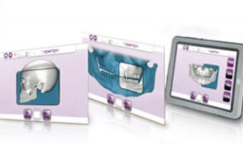
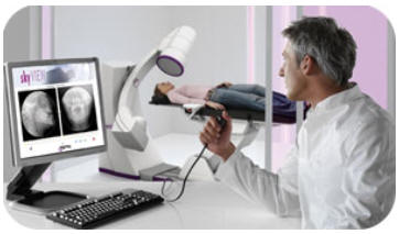
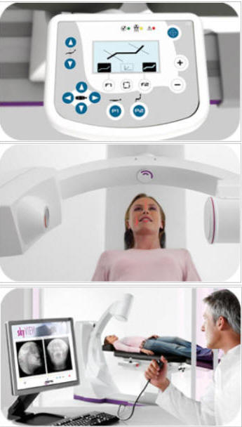
Virtual control panel
The virtual control
panel, which can be installed on a PC, allows
all diagnostic activities to be controlled from
a workstation.For example, the user can link up
a mini tablet-PC with touch screen to pilot the
machine while comfortably positioned outside the
X-ray area.Store images on a memory card or
share them over your local network through the
industry standard Ethernet.
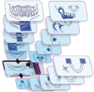
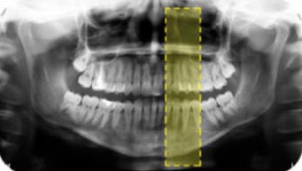
15 diagnostic programs
Standard
orthopanoramic or paediatric projections
with automatic selection of correct
morphology.
Frontal and lateral
views of the maxillary sinuses and
multi-angle lateral and posterior-anterior
views of the temporo-mandibular joint.
Partial projections
can be selected as a comfortable alternative
to intra-oral images for patients with a
strong gag reflex.
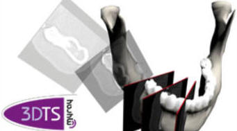
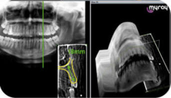
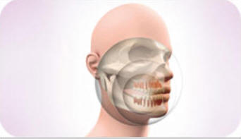
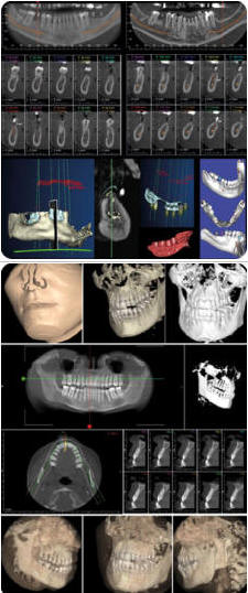
3DTS application
Hyperion’s additional
three-dimensional 3DTS application enables you
not only to view the panoramic image of the
dentition, but once the 3DTS process is
complete, you can also explore the third
dimension related to a specific volume of the
upper and lower jaw.
Inspection of surgical
site
The selection of a region
of interest is done within a rectangular area
directly on a panoramic radiographic image of
the patient in question, or by a template of an
average patient. Field of view: 4x4x10cm.
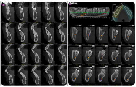
Being able to glance
through transversal slices of the area in
question on a PC screen is extremely useful to
whoever practices implantology, simply because
it offers accurate radiographic data to work
with, perfect for reliable (1:1) measurements,
with the precision of 0.15mm pixel size.
SkyView combines a cone beam X-ray
source with a latest generation image intensifier in order
to deliver high-definition 3D images with low X-ray doses.
With SkyView you can
obtain three-dimensional reconstructions of
teeth and the entire maxillofacial area.High-resolution
images with a 9” or 6” field of view can be
completed with a close-up zoom of a 4” diameter
section.Images are completely free from
geometric deformation, perfectly measurable with
micrometric precision and reliable over time.
Safety and wellbeing
The high sensitivity of
the image intensifier, the scan speed and the
pulsed X-ray emission system ensure that X-ray
doses are very low indeed.A natural, unhindered
position means no claustrophobia or anxiety.
With SkyView, the scanning experience is a
pleasant one.SkyView can easily be installed in
any dental surgery: X-ray screening requirements
are comparable to those of a panoramic system.
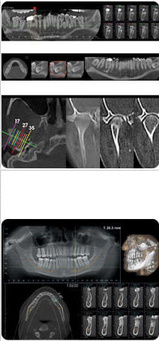
Laser beam positioning
and Scout method
The software-guided Scout
procedure is extremely high-precision and
involves the acquisition of two preview (Scout)
X-ray images at extremely low dosages; these
previews are used to identify the centre of
volumetric reconstruction.
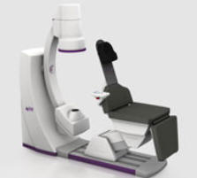
Above: New reclining chair for easy access
The new reclining patient
chair is automatically lowered into the access
position to make seating the patient easier; the
patient’s head is supported by the comfortable
headrest, a soft height-adjustable cushion
designed to stabilise the cranium; then the
chair stretches out to perform the quick X-ray
examination.
Image quality and
precision
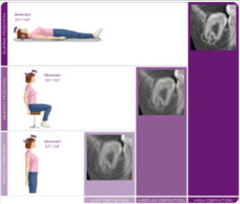
The supine position gives
the best possible degree of immobility without
having to resort to constriction of the cranium,
thus ensuring naturally enhanced image quality
Thanks to Isotropic Voxels, measurements are
reliable and on a 1:1 scale whatever the
reference plane of the measured sections..
Simple, yet effective positioning procedures
ensure the X-ray will never have to be repeated
because of alignment errors.
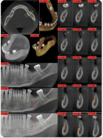
Perfect data
presentation
At the end of the
acquisition/volumetric reconstruction procedure,
the dentist will have multiple views of the
patient’s anatomy: · a curvilinear cross-section
that resembles the “panoramic” image· a moveable
3-D model· a coronal cross-section that allows
exploration of the upper/lower dental arches·
ten transverse cross-sections that are
especially useful for the linear and angular
measurements so often used with implants.